Figure 2.

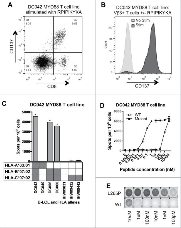
MYD88 L265P is recognized by an HLA-B*07:02-restricted CD8+ T cell line. (A, B) The MYD88L265P RPIPIKYKA-specific T cell line was stimulated overnight with or without peptide (10 μM) and then analyzed by flow cytometry. (A) Peptide stimulated cells were gated on single lymphocytes and assessed for expression of CD8+ and CD137. This showed that activated (CD137+) cells were CD8+. (B) After overnight incubation with or without peptide, cells were gated on single lymphocytes and then on expression of CD8+ and TCR-Vβ3. Stimulated CD8+ TCRVβ3+ cells express CD137, demonstrating that the relevant TCR is from the Vβ3 family. (C) IFNγ ELISPOT results from an HLA restriction experiment showing that the RPIPIKYKA peptide is presented in the context of HLA-B*07:02. T cells (1 × 10⁵ cells) were incubated with a panel of LCL (2 × 10⁵ cells/well) that were matched with the donor T cells at 0–2 alleles and that had been peptide-pulsed with RPIPIKYKA (10 μM). Background spot numbers (T cells plus LCL without peptide pulse) were subtracted. * = TNTC. Gray boxes indicate alleles present in each donor. (D) ELISPOT data showing recognition of MYD88L265P RPIPIKYKA (solid circles) at over 100,000-fold lower peptide concentration than MYD88WT RLIPIKYKA (open circles). Data points at the top of the curve are an underestimate, as spots were TNTC. (E) A subset of ELISPOT wells from the experiment described in Fig. 2D.
