
Figure 9.
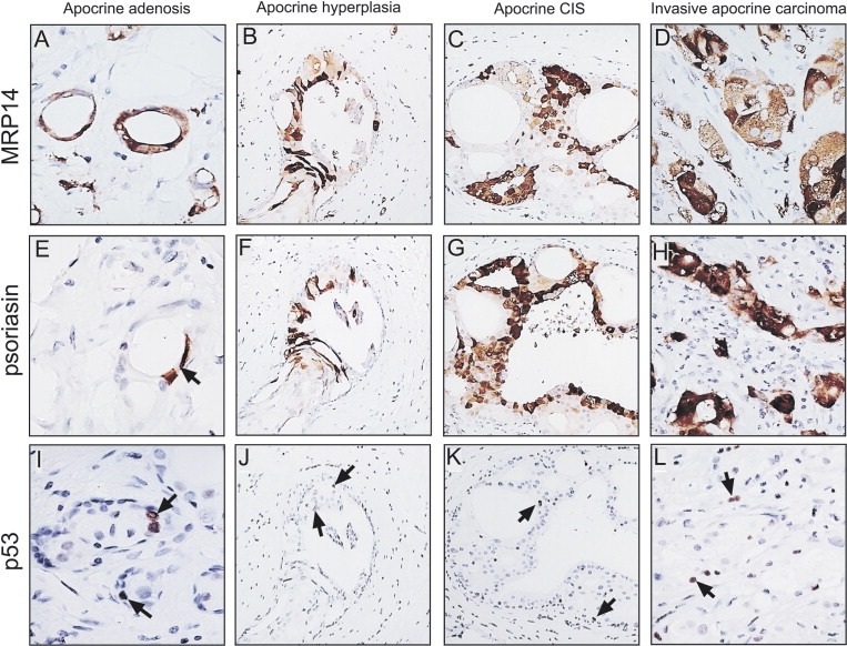
IHC staining of an invasive apocrine carcinoma using antibodies against components of the benign apocrine signature. (A, E, and I) Area with AA stained with antibodies against (A) MRP14, (E) psoriasin and (I) p53. (B, F, and J) Area with apocrine hyperplasia stained with antibodies against (B) MRP14, (F) psoriasin, and (J) p53. Area with apocrine CIS (cribriform) stained with antibodies against (C) MRP14, (G) psoriasin, and (K) p53. (D, H, and L) Area with invasive disease stained with antibodies against (D) MRP14, (H) psoriasin and (L) p53. The antibodies were used at the dilutions given in the legend of Figure 3.