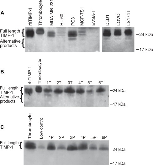
Figure 2.

Detection of TIMP‐1 and alternative TIMP‐1 products in cancer cell lines (A), lysates of tumor samples from CRC patients (B) and in plasma from CRC patients (C). Equal amounts of protein were separated by SDS‐PAGE, and TIMP‐1 was subsequently detected with a monoclonal anti‐TIMP‐1 antibody (VT7). Low control, platelet lysate (contains high TIMP‐1 protein levels) and recombinant human (rh) TIMP‐1 serve as controls. The plasma samples are representative of a total of 12 samples analysed. The experiment was repeated once with similar results.
