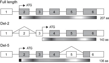
Figure 5.

Schematic representation of full‐length and alternatively spliced TIMP‐1. Exons are numbered from 1 to 6 and translated regions are shown in grey. White boxes indicate untranslated regions. Translation initiation sites are marked by black arrow and ATG. The corresponding translated proteins are shown underneath the mRNAs and the theoretical size is marked with number of amino acids (aa). The signal peptide sequences are shown as black boxes at the N‐terminal end of full‐length TIMP‐1 and del‐5 variant. The translation initiation site in del‐2 TIMP‐1 is shifted from exon 2 to exon 3, which results in a shorter protein lacking the signal peptide. A theoretical translation of the del‐5 variant results in a protein consisting of 136 aa due a shift in reading frame of the exon 6 sequence.
