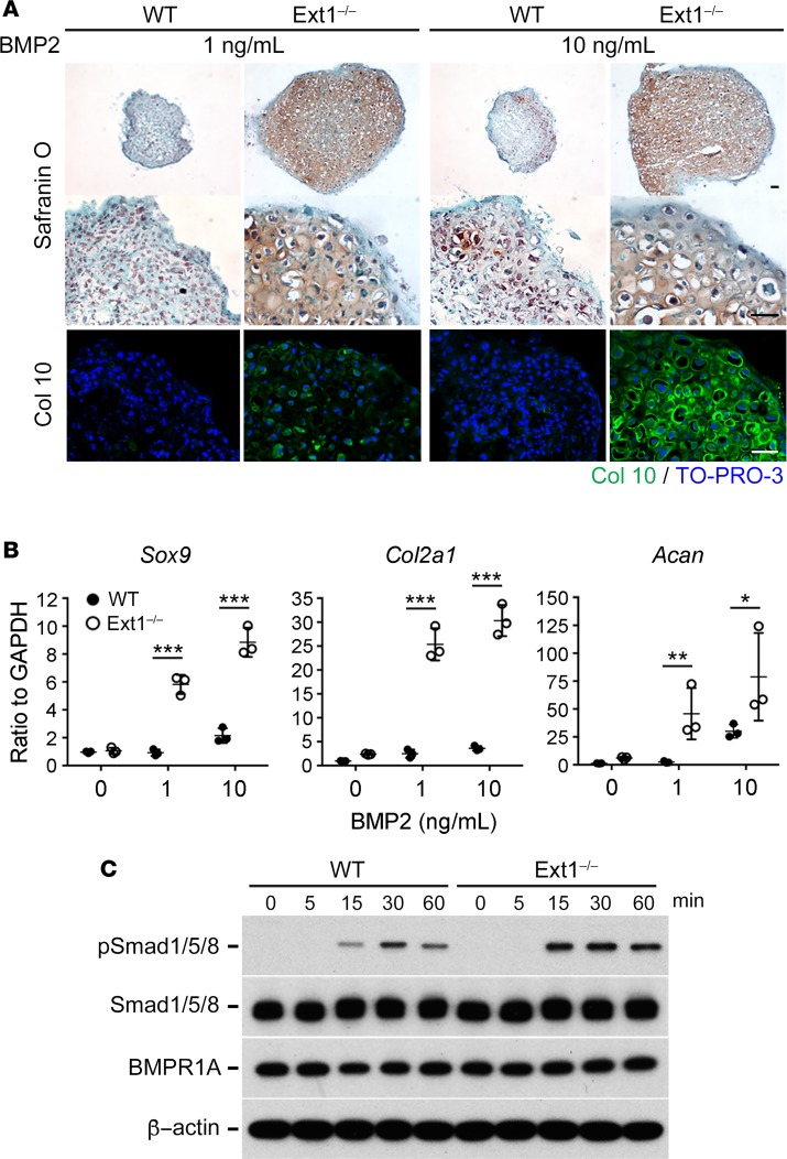
Figure 3. Ext1-deficient perichondrium-derived mesenchymal progenitor cells (PDPCs) exhibit enhanced chondrogenic differentiation and BMP signaling.
(A and B) Ext1-deficient (Ext1–/–) and WT (Ext1flox/flox; WT) PDPCs were cultured as 3-dimensional cell pellets for 2 weeks in the presence of 1 ng/ml or 10 ng/ml of BMP2. (A) Safranin O staining and immunohistochemical staining of type X collagen (Col10) of pellets. Pellets derived from Ext1-deficient PDPCs are larger and more intensely stained with Safranin O and for type X collagen than those derived from WT PDPCs. Scale bars: 0.1 mm. Data shown are representative of 3 independent experiments. (B) Expression of chondrogenic markers in pellet cultures derived from Ext1-deficient (Ext1–/–) and WT (Ext1flox/flox; WT) PDPCs. Expression of Sox9, Col2a1, and Aggrecan (Acan) was evaluated by qPCR. Gapdh was used as an internal control for normalization. Means ± SD (n = 3) are shown as horizontal bars. P values were determined by two-way ANOVA. *P < 0.05, **P < 0.01, ***P < 0.001. (C) Time course of BMP2-induced phosphorylation of Smad1/5/8. Ext1-deficient and WT PDPCs in monolayer cultures were stimulated with 10 ng/ml BMP2, and cells were lysed after indicated time of incubation. Cell lysates were immunoblotted with antibodies to phosphorylated Smad1/5/8, total Smad1/5/8, BMPR1A, and β-actin. This experiment was performed 3 times with similar results.

