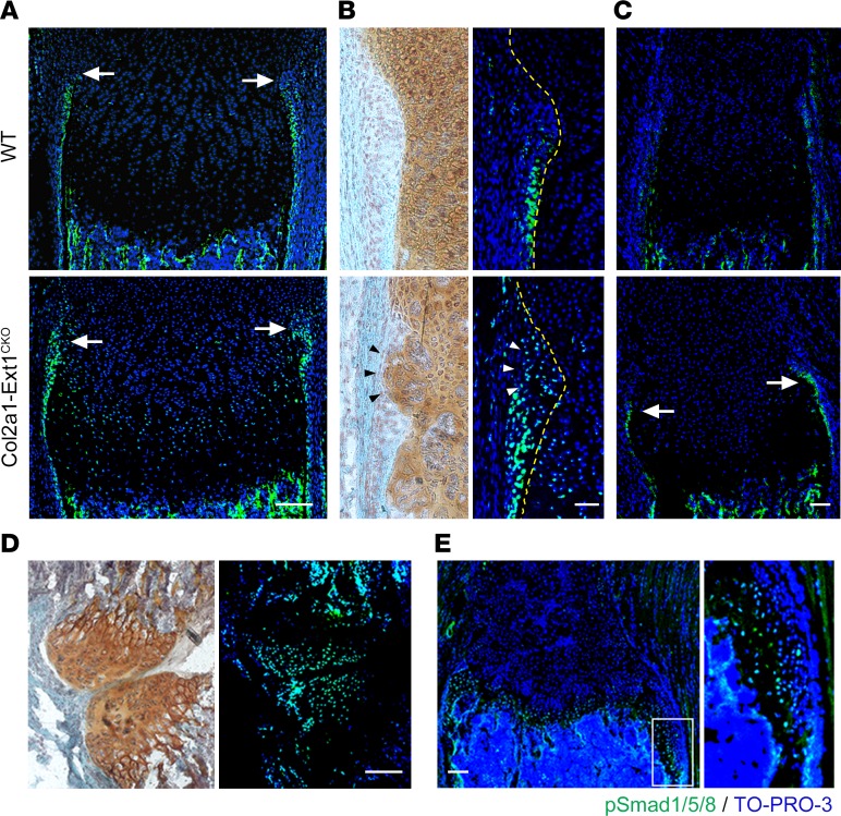
Figure 4. BMP signaling is upregulated in the perichondrium and osteochondromas of Col2a1-Ext1CKO mice.
(A–C) Frozen sections of the radius (A, B) and a rib bone (C) from P10 Col2a1-Ext1CKO and control (Ext1flox/flox; WT) littermates were double-labeled with anti-pSmad1/5/8 antibody (green) and TO-PRO-3 (blue) or stained with Safranin O. (A) pSmad1/5/8 expression in the perichondrium of the radius. The location of the groove of Ranvier is indicated by arrows. Note that the intensity and spatial extension of pSmad1/5/8 immunoreactivity is increased in the perichondrium of Col2a1-Ext1CKO mice. (B) High-power views of the groove of Ranvier. In control mice (Ext1flox/flox), most pSmad1/5/8 immunoreactivity is seen in the diaphyseal side of the groove of Ranvier, and few pSmad1/5/8-immunoreactive cells are present within the groove per se. In Col2a1-Ext1CKO mice, the distribution of pSmad1/5/8-expressing cells is expanded both laterally and longitudinally. The cells forming the abnormal cell cluster within the groove of Ranvier (indicated by arrowheads) are also pSmad1/5/8-positive. Broken lines depict the perichondrium/growth plate boundary. (C) pSmad1/5/8 expression in the perichondrium of a rib bone. While WT perichondrium shows little pSmad1/5/8 immunoreactivity, perichondrium of Col2a1-Ext1CKO mice displays strong pSmad1/5/8 immunoreactivity (arrows). (D) pSmad1/5/8 expression in osteochondromas formed in the forelimb of P28 Fsp1-Ext1CKO mouse. Adjacent sections were stained with Safranin O (left panel) or double-labeled with anti-pSmad1/5/8 antibody and TO-PRO-3 (right panel). Cells forming the cartilage cap of osteochondromas are pSmad1/5/8-positive. (E) pSmad1/5/8 expression in overgrown cartilage in a rib bone of P28 Fsp1-Ext1CKO mouse. The image on the right shows an enlarged view of the area indicated by a rectangle. Cells forming overgrown cartilage are immunoreactive to pSmad1/5/8. Scale bars: 0.1 mm (A, C, D, E); 20 μm (B). Data shown are representative images; each analysis was performed on at least 3 mice per genotype.

