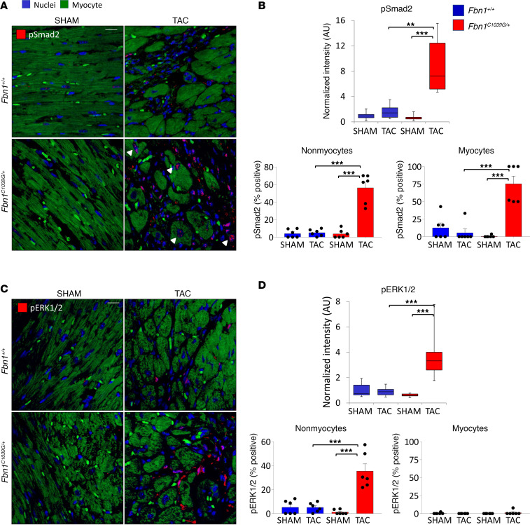
Figure 3. Smad2 and ERK1/2 activation are differentially increased in the myocyte and nonmyocyte compartments of failing Fbn1C1039G/+ hearts.
(A) Representative sections of pSmad2 (red) immunostaining. Scale bar: 20 μm. White arrowheads point to examples of positively stained myocyte nuclei. (B) Summary quantification of total intensity normalized to Fbn1+/+:sham and labeling index (percentage positive cells) of pSmad2 in the nonmyocyte and myocyte compartments. (C and D) Representative sections and summary quantification of pERK1/2 (red) immunostaining. Scale bar: 20 μm. n = 6 fields per group. **P < 0.01, ***P < 0.001, 1-way ANOVA, Tukey’s correction.

