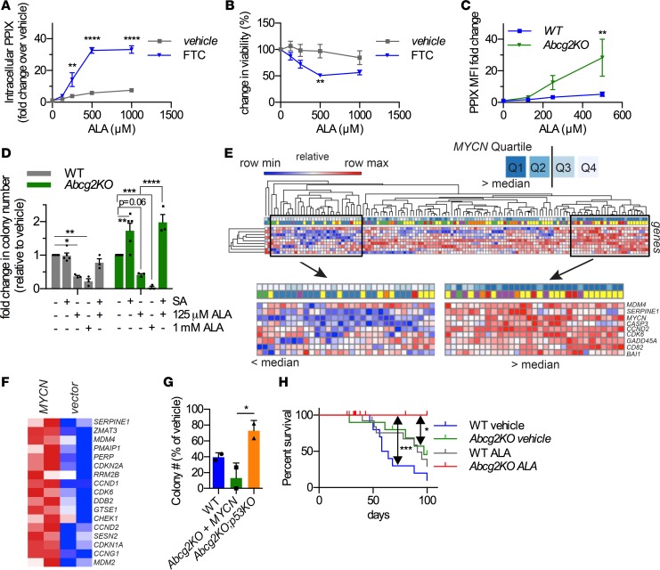
Figure 4. Porphyrin cytotoxicity is abrogated by p53 absence, and AML is cured by enhancing porphyrins in the absence of ABCG2.
WT-MYCN HPCs were incubated with increasing doses of ALA for 72 hours in the absence or presence of FTC. (A) Cell viability and (B) PPIX levels were measured (n = 3, mean ± SEM). (C) Abcg2-KO MYCN-HPCs accumulate high levels of porphyrins compared with WT-MYCN HPCs (n = 3, mean ± SEM). (D) High porphyrin levels are especially detrimental to colony formation of Abcg2-KO MYCN-HPCs (n = 4, mean ± SEM). (E) AML patients with MYCN levels in the upper quartile display strong activation of a p53 gene set. (F) Abcg2-KO MYCN-HPCs show activation of p53 target genes. (G) CFU-c assays were performed using MYCN-transduced HPCs isolated from mice of the indicated genotypes. Cells were incubated with ALA and colonies were counted. Loss of p53 renders Abcg2-KO MYCN cells resistant to porphyrin toxicity (2 independent experiments, with 2 replicates per condition per experiment, and values shown as an aggregate mean ± SD; 1-way ANOVA). (H) WT or Abcg2-KO MYCN-expressing HPCs were treated with ALA for 24 hours prior to transplantation into lethally irradiated recipient mice. Survival curve showing that Abcg2-KO cells treated with ALA did not produce leukemia. Median survival for WT vehicle, Abcg2-KO vehicle, and WT ALA were 61 days, 97 days, and 91 days, respectively. The Abcg2-KO and WT leukemia were myeloperoxidase positive, as shown in Supplemental Figure 6. Median survival for Abcg2-KO ALA was not determined (n = 10 for vehicle-treated and n = 14 for ALA-treated samples; P = 0.05 between WT vehicle and Abcg2-KO vehicle, log-rank Mantel-Cox test). *P < 0.05, **P < 0.01, ***P < 0.001, ****P < 0.0001. Two-way ANOVA was used for A–D.

