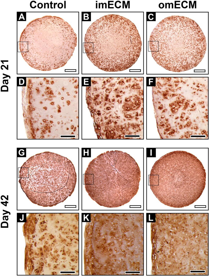
Fig. 5.

Immunohistochemical staining of collagen type II in 3D MSC-seeded GelMA constructs. (A–F) Constructs on day 21; (A–C) Low magnification, scale bar = 1 mm; Area of magnification shown by black box; (D–F) High magnification, scale bar = 200 μm. (G–L) Constructs on day 42; (G–I) Low magnification, scale bar = 1 mm; Area of magnification shown by black box; (J–L) High magnification, scale bar = 200 μm.
