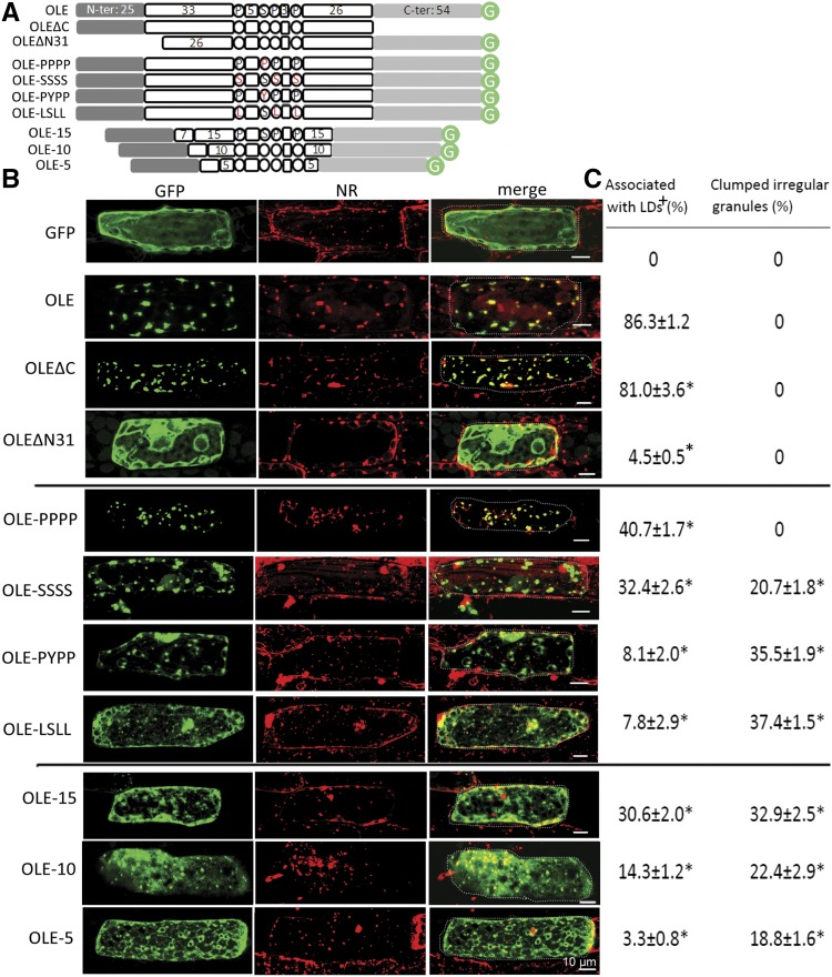
Figure 2.
Subcellular localization of native and recombinant oleosins in P. patens cells after transient gene expression. A, In linear portions, the native and recombinant oleosins of P. patens (OLE). The N- and C-terminal portions are amphipathic and are shown in shaded boxes. The whole hairpin of ∼72 residues is hydrophobic; its loop (PX5SPX3P) and the two arms (33 and 26 residues) are in white circles or boxes. Three sets of recombinant oleosins include those with deletion of the C- or N-terminal portion, alteration (highlighted in red) of the four completely conserved P, S, P, P in the loop, and reduction of the hairpin length. In the lowest subpanel, the boxed 7 represents the initial 7 residues of the hairpin arm required for ER targeting. Circled G represents GFP. B, Images of individual cells after transient expression of the respective DNA constructs encoding GFP alone or native/recombinant oleosin with its C terminus attached to GFP. Cells were transformed with the DNA constructs via bombardment, and after 12 h, GFP fluorescence and Nile Red (NR) staining of LDs were monitored with CLSM. In the merge images, a dotted line outlines the cell circumference. Bars = 10 μm. C, Quantification of fluorescence in different subcellular locations with ImageJ. + indicates the proportion of GFP associated with LDs (stained with Nile Red) and irregular granules (determined with CLSM). The remaining proportion was mainly with cytosol (in the test of OLEΔN31) or ER (in the tests of other recombinant OLE); the proportion in these other subcellular locations could not be assigned precisely and are not shown. *P < 0.05 compared with OLE by Student’s t test.

