Figure 4.
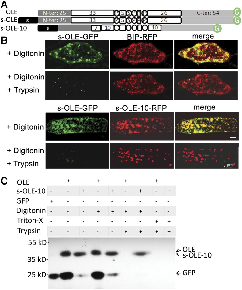
Subcellular localization of recombinant oleosins, with emphasis on their association with the luminal or cytosolic side of ER, in P. patens cells after transient gene expression or tobacco BY2 cells after stable transformation. A, In linear portions (following those described in Fig. 2 legend), OLE, s-OLE, and a hairpin-shortened oleosin (s-OLE-10) with an attached 21-resiude N-terminal ER-targeting peptide (s) of P. patens aspartic proteinase. B, Images of portions of a P. patens cell after transient expression of DNA constructs (s-OLE-GFP, s-OLE-10, BIP-RFP) and then subjected to fluorescence protease protection test. In the top portion, the cell was cotransformed with DNA constructs encoding s-OLE-GFP and BIP-RFP (ER-lumen marker). In the bottom portion, the cell was cotransformed with DNA constructs encoding s-OLE-GFP and s-OLE10-RFP. The transformed cells were permeated with digitonin and then digested with trypsin. GFP and RFP were monitored with CLSM. Bars = 5 μm. C, Immunoblotting of total extracts of tobacco cells after stable transformation with DNA constructs and then treatments with detergents and trypsin. In the top portion, each column shows the DNA construct (OLE-GFP, s-OLE-10, or GFP without OLE) used and the subsequent detergent (digitonin and Triton-X) and/or trypsin treatments. After SDS-PAGE, the gel was immunoblotted with antibodies against GFP. Positions of molecular-weight markers are on the left. Projected positions of OLE-GFP (42 kD), s-OLE-10-GFP (42 kD, barely separable from OLE-GFP), and GFP (27 kD) are on the right.

