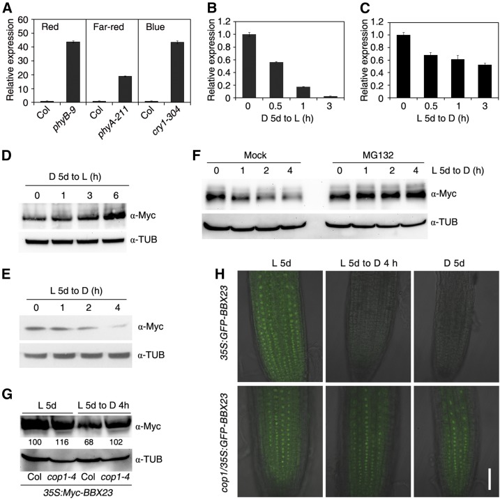
Figure 4.
BBX23 is regulated by light. A, BBX23 expression in different photoreceptor mutants. Col and phyB-9, phyA-211, and cry1-304 mutants were grown under continuous red, far-red, or blue light conditions, respectively, for 5 d. B, BBX23 expression during dark-to-light transition. Col wild-type seedlings were grown in darkness for 5 d followed by light exposure for 0.5, 1, and 3 h. C, BBX23 expression during light-to-dark transition. Col wild-type seedlings were grown in white light for 5 d followed by dark treatment for up to 3 h. For A to C, RT-qPCR was performed, and the relative expression levels were normalized to those of IPP2. Mean ± sd, n = 3. D, Immunoblotting of Myc-BBX23 during dark-to-light transition. The 35S:Myc-BBX23 (line #1) seedlings were grown in darkness for 5 d and then exposed to light for the indicated periods of time. E, Immunoblotting of Myc-BBX23 during light-to-dark transition. Five-day-old seedlings were transferred to darkness for the indicated periods of time. F, Immunoblotting of Myc-BBX23 during light-to-dark transition. Five-day-old light-grown 35S:Myc-BBX23 seedlings were incubated without (Mock) or with 50 µm MG132 and transferred to darkness for the indicated periods of time. G, Detection of Myc-BBX23 in the cop1 mutant. Five-day-old light-grown seedlings of 35S:Myc-BBX23 and cop1-4/35S:Myc-BBX23 were transferred to darkness for 4 h. Relative protein abundance of Myc-BBX23 are calculated and shown below the blotting bands. For D to G, blotting against a tubulin antibody serves as equal loading controls. H, GFP fluorescence. 35S:GFP-BBX23 and cop1-4/35S:GFP-BBX23 seedlings were grown in darkness and white light for 5 d, or white light-grown seedlings were then transferred to darkness for 4 h. GFP-BBX23 is localized in the nucleus of Arabidopsis root cells. Merged images of GFP fluorescence and DIC channels are shown. Bar = 50 µm. For B to H, D denotes dark and L indicates light.

