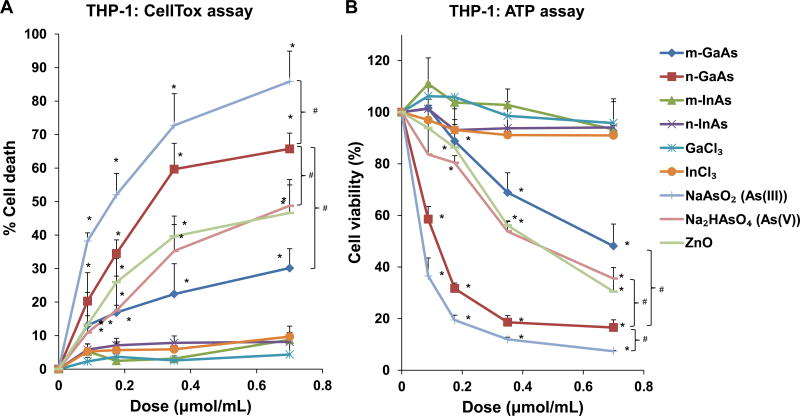
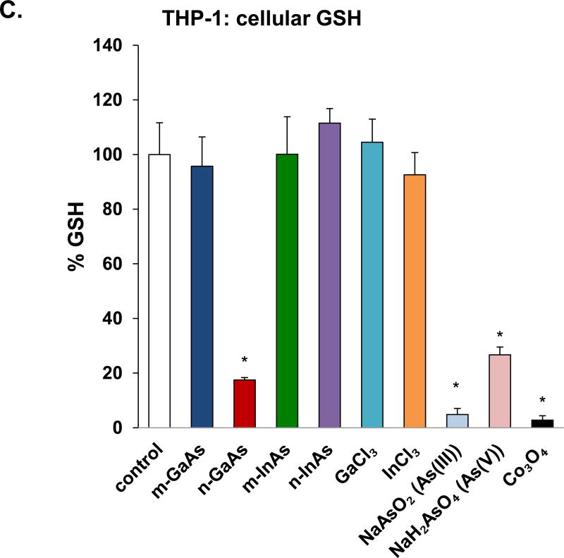
Figure 2.
Cell viability and oxidative stress analysis in THP-1 cells, following treatment with particulate and ionic materials. (A) Assessment of cell death by the CellTox assay. (B) Assessment of THP-1 viability by the ATP assay. The materials were added to the cell culture medium at 0.07–0.7 µmol/mL for 24 h. The corresponding mass doses are listed in Table 3. (*) p < 0.05 compared to control; (#) p < 0.05 compared to n-GaAs. (C) Intracellular GSH depletion was determined by a luminescence-based GSH-Glo kit. THP-1 cells were exposed to III-V materials at 0.35 µmol/mL (=50 µg/mL GaAs) for 24 h. GSH abundance was calculated as the fractional luminescence intensity of treated vs. untreated cells. The relative GSH abundance in non-treated cells was regarded as 1.0. (*) p < 0.05 compared to control.


