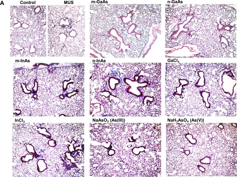
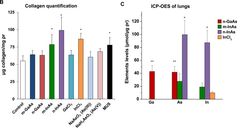
Figure 7.
Pro-fibrogenic effects and elemental analysis, 21 D after initial exposure to particulate and ionic III-V materials. (A) Visualization of collagen deposition (blue-color staining) in the lung by Masson’s trichrome staining (100× magnification). MUS served as a positive control. (B) Assessment of total collagen content by using a Sircol kit (Biocolor Ltd., Carrickfergus, UK). (*) p < 0.05 compared to control. (C) Elemental Ga, As and In content of the lung by ICP-OES, normalized for protein content in the lung tissue. The intact lungs were collected and processed for ICP-OES analysis as similar as Figure 4. None of the above III-V elements were detected in lung tissue 21 D after m-GaAs, GaCl3, As(III) and As(V) exposure. (**) p < 0.05 compared to m-GaAs. (#) p < 0.05 compared to m-InAs.


