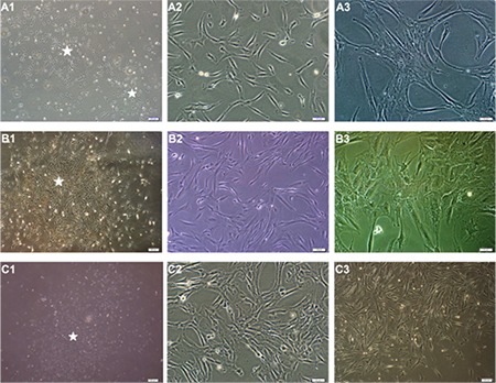
Figure 1. Isolated cells from human bone marrow- (A1-A3), human adipose tissue- (B1-B3), and human Wharton’s jelly- mesenchymal stem cells (MSCs) (C1-C3) distributed sparsely on the culture flasks displayed mostly fibroblast-like, spindle-shaped morphology during the early days of incubation. Small colonies (asterisks), called colony-forming units, appeared within 9-12 days (A1, A2: P0 - 9th day, B1: P0 - 12th day, C1: P0 - 12th day). These primary cells reached monolayer confluence within 15-17 days. In the later passages, most of these MSCs exhibited large, flattened, or fibroblast-like morphology (A3, C3: P3 - 3rd day, A2: P2 - 4th day, C2: P1 - 6th day, and B3: P3 - 7th day).

