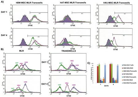
Figure 5. Inhibitory effect of human mesenchymal stem cells (hMSCs) on the proliferation of phytohemagglutinin (PHA)-CD3+ T cells as detected by carboxyfluorescein succinimidyl ester (CFSE). (A) Representative analysis of the altered rate of PHA-CD3+ T cells in the mixed lymphocyte reactions (MLR) and transwell experiments when co-cultured with hMSCs was performed by CFSE labeling at days 1 and 4. Solid purple histogram represents data of T cell-only culture, while green and pink peaks show the result of direct (MLR) and indirect (transwell) culture, respectively. (B) Comparative analyses of three cell cultures indicate that hBM-MSCs are the most effective cell type in the repression of induced T-cell proliferation. (C) Graphical representation of the cell proliferation in different cell cultures supports the results of previous data (n=3, mean ± standard deviation; Δ p<0.001). hBM: Human bone marrow, hAT: human adipose tissue, hWJ: human Wharton’s jelly.

