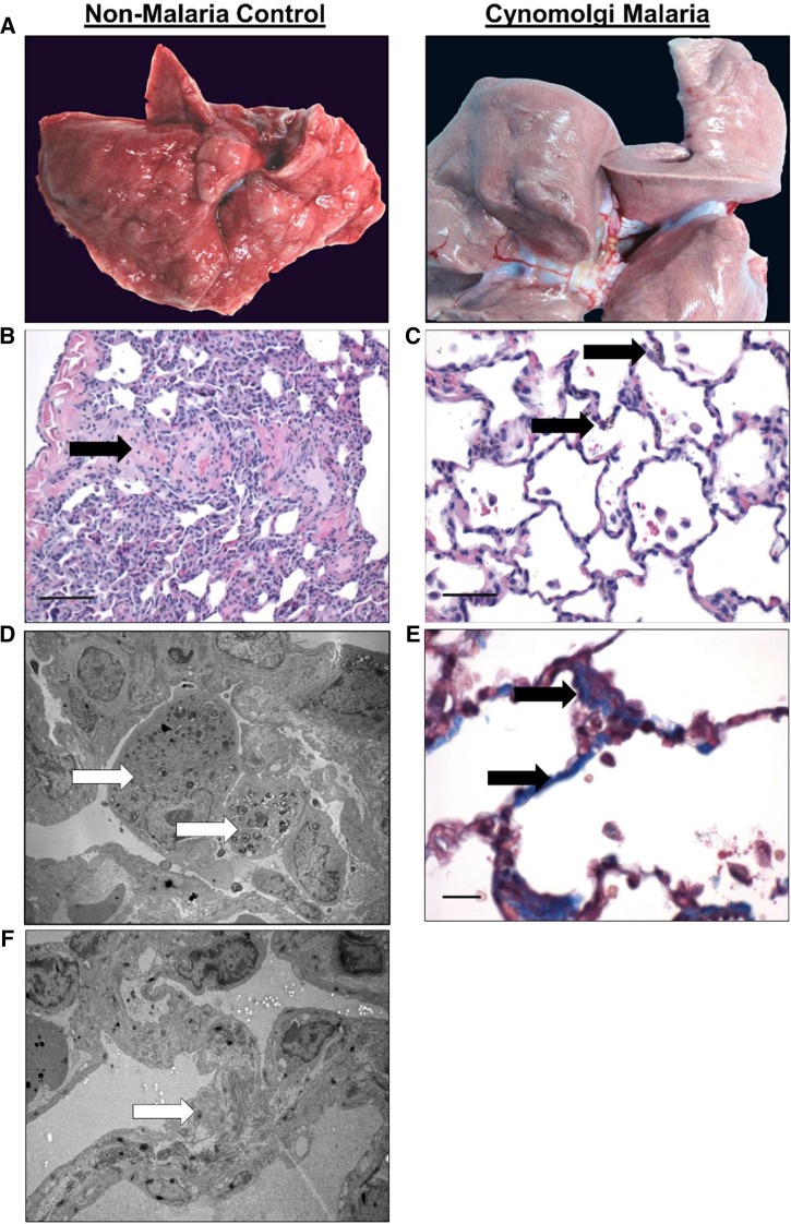
Figure 3.
Pulmonary lesions during severe and complicated cynomolgi malaria. (A) Comparison of a lung collected at necropsy from the rhesus macaque with severe and complicated cynomolgi malaria and a rhesus euthanized for reasons other than malaria. (B and C) Digital micrographs of sections from Panel A stained with hematoxylin and eosin. Black arrows indicate eosinophilic material in the parenchyma (B) and type II cell hyperplasia and alveolar macrophages (C). (D and F) Transmission electron micrographs from the infected lung in Panel A depicting a macrophage-containing hemozoin (D) and fibrils contained within type II cells (F). (E) Lung tissue section stained with Masson’s trichrome blue staining. Arrows indicate increased deposition of collagen within the interstitium in the lung.

