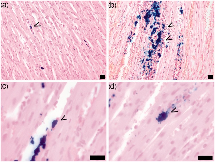
Fig. 3.
Number, localization, and morphology of BMMNCs in the myocardium. The number of BMMNCs (arrows) in the myocardium was qualitatively evaluated as: 0 = no cells, 1 = a few cells (a), or 3 = a large amount of cells (b). The BMMNCs mainly localized in slit-like spaces within myocyte bundles or inside vessels or remained in aggregates. (c) Single elongated and flattened cells were situated along the capillary endothelial cells and myocytes in a parallel fashion (arrow). (d) Single cells were also detected among the myocytes (arrow). Prussian blue staining. Scale bar = 20 µm.

