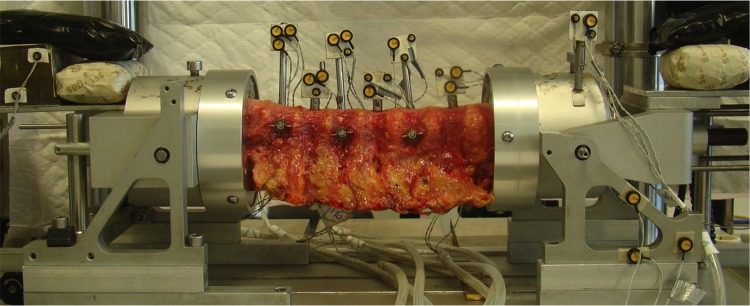
Figure 4.
The experimental setup is shown with a human cadaveric thoracic spinal specimen positioned in the spinal motion simulator. The proximal and distal vertebrae were potted in a casting-mold and partially buried in a low melting point (48°C) bismuth alloy (Cerrolow-147; 48.0% bismuth, 25.6% lead, 12.0% tin, 9.6% cadmium, and 4.0% indium). The proximal and distal vertebrae were fixed securely into the alloy by adding screws into the vertebral body. All articulating parts were kept free. During the experiment, the spinal specimens were kept moist with 0.9% saline spray. A materials testing machine applied loads to the setup to induce flexion-extension, lateral bending and axial rotation. The markers with the LEDs were rigidly fixed to the vertebrae. An Optotrak camera registered the movement of the infrared-emitting LEDs.

