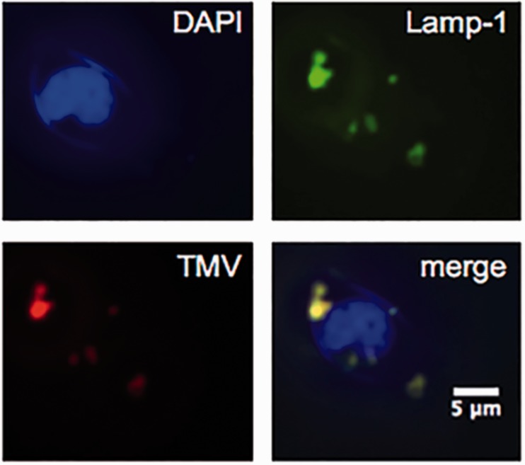
Figure 3.
Karpas 299 cell interactions with TMV. Nuclei were stained with DAPI (blue), endolysosomes were stained with mouse anti-human Lamp-1 antibody followed by secondary Alexa Fluor 488 goat anti-mouse antibody (green), and TMV particles were stained with primary rabbit anti-TMV antibody followed by secondary Alexa Fluor 555 goat anti-rabbit antibody (red). Merging of all three channels indicated co-localization of TMV with the endolysosomes (yellow). (A color version of this figure is available in the online journal.)
DAPI: 4',6-diamidino-2-phenylindole; TMV: tobacco mosaic virus

