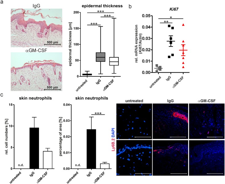
Fig 2. Neutralization of GM-CSF ameliorates cellular changes associated with IMQ-induced psoriasis.
Psoriasis was induced by topical application of IMQ in wild-type mice treated with isotype control (IgG) or a GM-CSF neutralizing antibody (αGM-CSF); mice were sacrificed on day 6. Untreated animals were not treated with IMQ. (a) Representative hematoxylin and eosin (HE) staining of back skin and quantification of epidermal thickness of HE-stained samples (untreated n = 3; IgG n = 5; αGM-CSF n = 6). (b) Ki67 mRNA expression (right: untreated n = 4; IgG n = 6; αGM-CSF n = 7; each symbol represents the quantified measured mRNA of one mouse) in skin by qPCR. (c) Quantification of neutrophil distribution by flow cytometry (left: n = 4) and by immunofluorescence staining with Ly6B.2 (middle: untreated n = 4; IgG n = 8; αGM-CSF n = 11) based on representative staining patterns (right). Neutrophils were not detectable (n.d.) in untreated mice. Scale bars equal 100 μm. Graphs show mean values ± SEM, except for right panel of (a) (Box-Whisker-Plot of epidermal thickness). *p-value<0.05, **p-value<0.01, ***p-value< 0.001 as determined by Mann-Whitney-U-Test.

