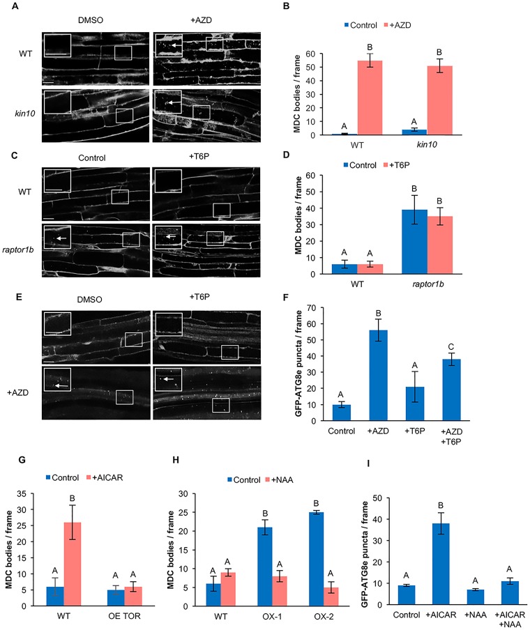
Fig 5. SnRK1 acts upstream of TOR in the autophagy pathway.
(A) WT and kin10 seedlings were grown on ½ MS plates for 7 days. Seedlings were transferred to ½ MS liquid medium supplemented with 10 μM AZD or DMSO for 3 hours, followed by MDC staining. Confocal microscopy was used to visualize autophagosomes (white arrows) in roots. The insets show enlargements of the indicated boxes. Scale bars = 20 μm. (B) Quantification of autophagy activity as shown in (A). Upon inhibition of TOR with AZD, autophagy was still activated in kin10 mutant seedlings. (C) WT and raptor1b seedlings were grown on ½ MS plates for 7 days. Seedlings were transferred to ½ MS liquid medium supplemented with 0.1 mM T6P for 3 hours, followed by MDC staining. Confocal microscopy was used to visualize autophagosomes (white arrows) in roots. The insets show enlargements of the indicated boxes. Scale bars = 20 μm. (D) Quantification of autophagy activity in (C). Upon inhibition of SnRK1 with T6P, autophagy activity was not affected in raptor1b seedlings. (E) GFP-ATG8e seedlings were grown on ½ MS plates for 7 days. Seedlings were transferred to ½ MS liquid medium supplemented with 0.1 mM T6P or 10 μM AZD or T6P plus AZD for 3 hours. Confocal microscopy was used to visualize autophagosomes (white arrows) in roots. The insets show enlargements of the indicated boxes. Scale bars = 20 μm (F) Quantification of autophagosomes labeled with GFP-ATG8e in (E). Upon inhibition of both TOR and SnRK1, autophagy was activated. (G) WT and OE TOR seedlings were grown on ½ MS plates for 7 days. Seedlings were transferred to ½ MS liquid medium supplemented with 10 mM AICAR for 1 hour, followed by MDC staining, and autophagosomes counted. Overexpression of TOR was able to suppress AICAR-induced autophagy. (H) WT, KIN10 OX-1 and KIN10 OX-2 seedlings were grown on ½ MS plates for 7 days. Seedlings were transferred to ½ MS liquid medium supplemented with 20 mM NAA or DMSO for 6 hours, stained with MDC and autophagosomes counted. Activation of TOR by auxin inhibited the constitutive autophagy in KIN10 overexpression lines. (I) Seven-day-old GFP-ATG8e seedlings were transferred to ½ MS liquid medium supplemented with 10 mM AICAR or 20 nM NAA or both AICAR and NAA. Activation of TOR by NAA blocked induction of autophagy by AICAR. For all graphs, different letters denote statistical significance for three biological replicates with at least 10 frames per replicate, p<0.05, t-test. Error bars indicate standard error.

