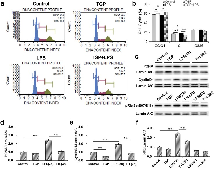
Fig 1. TGP inhibits cell proliferation of LPS-stimulated PC-3 cells.
PC-3 cells were incubated with LPS (2.0 μg/mL) in absence or presence of TGP (312.5 μg/mL). Cells were collected at 6 hr after LPS. (a) Cell cycle was measured using Muse™ Cell Cycle Kit. (b) Cell quantification of each cell cycle phase. (c-f) Cells were collected at 3 hr and 6 hr after LPS. (c) A representative gel for PCNA (upper panel), CyclinD1 (middle panel) and pRb (lower panel) was shown. (d) PCNA/Lamin A/C, (e) CyclinD1/Lamin A/C and (f) pRb/Lamin A/C. All experiments were repeated for three times. Data were expressed as means ± S.E.M. (N = 3), *P<0.05, **P<0.01.

