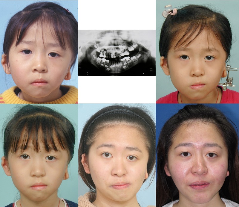
Fig 8. A female patient with Type IIB hemifacial microsomia involving the left side of face (case 13).
She was followed up at 5 years of age (above left and middle). The panorex X-ray showed hypoplastic and inferiorly displaced condyle without adequate glenoid fossa. She was 7 years 9 months of age during the distraction osteogenesis (above right). Facial appearance was improved at 8 years 6 months of age (below left). Significant facial asymmetry was noted at 21 years 1 month of age (below middle). She received two stages of surgical correction. The facial appearance was improved at 22 years 2 months of age (below right).

