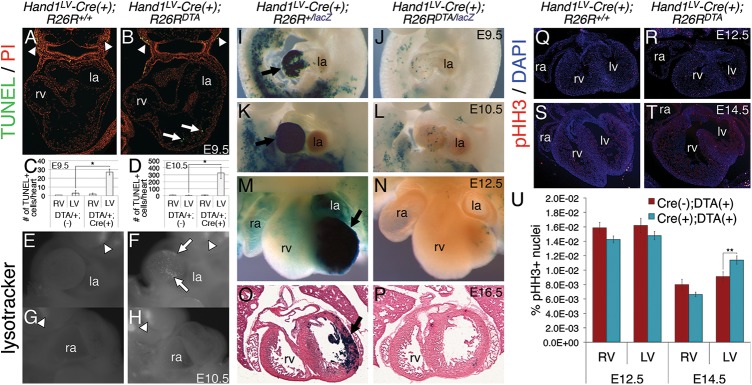
Fig 2. Hand1LV-Cre-mediated DTA activation results in LV cardiomyocyte cell death and hypoplastic LV, which, through cardiomyocyte proliferation, recovers fully by E16.5.
A, B) TUNEL on E9.5 sections of Hand1LV-Cre(+);R26RlacZ/+ control (A) and Hand1LV-Cre(+);R26RlacZ/DTA embryos. Sections are counterstained with propidium iodide (PI). C, D) Quantification of the number of TUNEL-positive cells per heart in control and Hand1LV-Cre(+);R26RlacZ/DTA embryos at E9.5 (C) and E10.5 (D). E-H) Whole mount lysotracker staining detecting cell death confirms that conditional activation of R26ReGFPDTA/+ via intercross to Hand1LV-Cre causes pronounced LV cardiomyocyte death as assayed at E10.5 (white arrow; n = 3). White arrows denote TUNEL or lysotracker-positive cells in the LV. White arrowheads denote TUNEL or lysotracker-positive cells in the pharyngeal arches. I-P) X-Gal staining to detect the R26RlacZ reporter allele demonstrates that activation of the R26ReGFPDTA allele nearly completely ablates the Hand1LV-Cre lineage (black arrow) by E9.5 (I, J; n = 5); however, LV hypoplasia is not apparent until E10.5 (K, L; n = 3) and persists at E12.5 (M, N; n = 4). Despite DTA-mediated ablation of the Hand1LV-Cre lineage, by E16.5, the initially hypoplastic LV shows a pronounced recovery, and LV size is comparable to controls (O, P; n = 3). Q-T) Immunohistochemistry for pHH3 at E12.5 (Q, R) and E14.5 (S, T) in Hand1LV-Cre(+);R26R+/+ control (Q, S) and Hand1LV-Cre(+);R26R+/DTA embryos (R, T). U) Quantification of pHH3+ cells relative to the number of DAPI+ pixels show that proliferation is not altered at E12.5, but is elevated specifically within the LV at E14.5. Data are represented as mean ± standard error of mean. Asterisks denote significance (p ≤ 0.05) as determined by student’s t-test.

