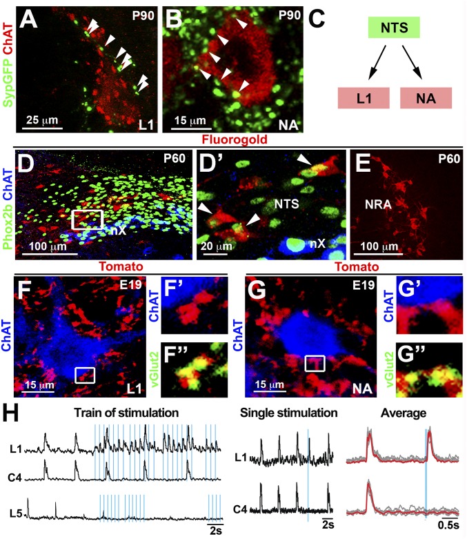
Fig. 4.
NTS neurons innervate expiratory and laryngeal motor neurons in newborn mice. (A and B) Injection of AAV encoding SypGFP into the NTS (n = 3) at P60 demonstrating the presence of GFP+ synaptic boutons (arrowheads) on L1 spinal (A) and nucleus ambiguus (NA) (B) motor neurons (ChAT+; red; single motor neurons are shown (Fig. S5 B and C). (C) Scheme illustrating that premotor neurons of the NTS connect to laryngeal and expiratory motor neurons. (D and D′) After injection of Fluorogold into the ventral L1 spinal cord, Fluorogold+ (red; arrowheads) neurons were present in the caudal NTS. Phox2b (green) and ChAT (blue) were used to distinguish Phox2b+ NTS neurons from Phox2b+/ChAT+ vagal motor nucleus (nX) on transverse sections. D′ shows a magnification of the boxed area in D. (E) Fluorogold+ cells (red) in the nucleus retroambiguus. (F and G) Intersectional strategy (Olig3creERT2/+;Phox2bFlpo/+;Ai65−/+ mice) to specifically label NTS axons and their synaptic terminals with Tomato fluorescent protein (Fig. S6). ChAT+ (blue) motor neurons at L1 spinal cord levels (F) and in the nucleus ambiguus (G) display numerous Tomato+ (red)/vGluT2+ (green) contacts. F′ and F″ and G′ and G″ show magnifications of the boxed areas in F and G, respectively. Single motor neurons are shown in F and G (overview provided in Fig. S6 C and D). (H, Left) Recordings of L1 (expiratory), C4 (inspiratory), and L5 (locomotion) motor roots in hindbrain-spinal cord preparations (n = 4) after trains of light stimulation (blue lines) on channelrhodopsin+ NTS cells. Light triggered only L1 responses, and not C4 or L5 responses. Note that inspiratory (C4) and expiratory motor roots (L1) are rhythmically active and fire synchronously in such preparations. (H, Middle) An evoked burst of L1 but not C4 motor root activity after a single light stimulation of the NTS. (H, Right) Superimposition of individual (gray) and average (red) traces of L1 and C4 recordings.

