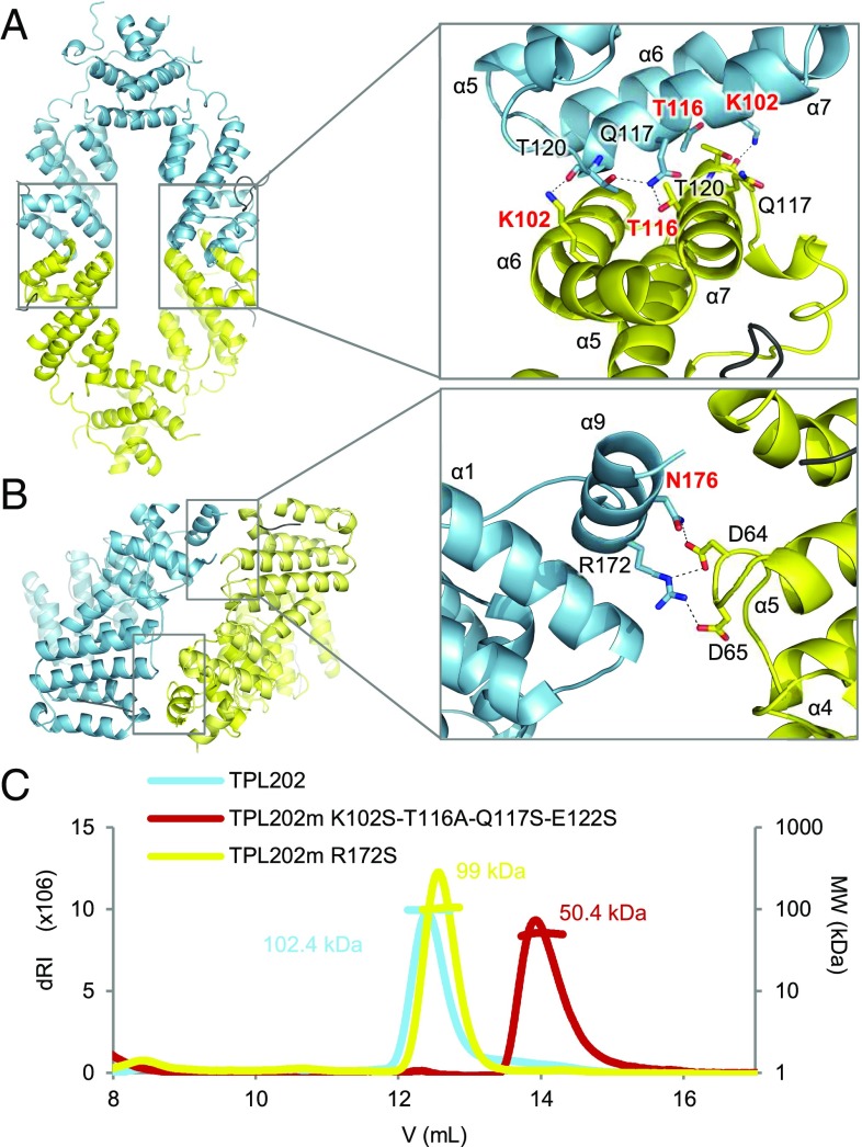
Fig. 2.
TPL tetramerization. (A) AtTPL184 tetrameric form I (dimers in blue and yellow). (Inset) Close-up view of the tetramerization interface with a rotation of about 90° to better view interacting residues. The interactions between chains C and D (Left) are slightly different from those between A and B (Right), as indicated in SI Appendix, Table S3. Residues mutated in different experiments are shown in red. (B) AtTPL184 tetrameric form II and close-up view of the interface. Main residues shown as sticks; distances <3.4 Å shown as dashes. (C) SEC-MALLS on TPL202 (blue) and TPL202 tetramerization mutants in both tetramerization interfaces I (red) and II (yellow). dRI, differential refractive index; MW, molecular weight.

