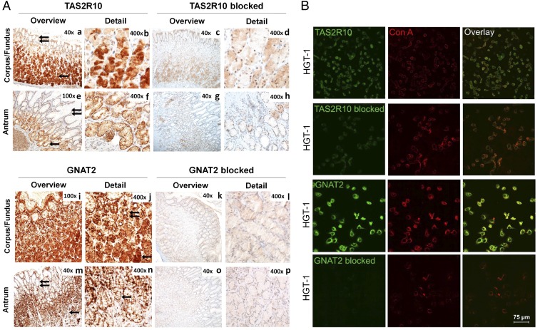
Fig. 3.
(A, a–p) Immunochemical localization of TAS2R10 and GNAT2 in (A) gastric tissue and (B) HGT-1 cells with and without preincubation with a blocking peptide. (a) In the gastric corpus/fundus, cytoplasmic reactivity of TAS2R10 in parietal and chief cells (one arrow) was detected whereas foveolar cells were negative (two arrows). Detail (b) shows parietal and chief cells. In the gastric antrum (e and f), very faint cytoplasmic and focal membranous reactivity of TAS2R10 in glandular cells was detected (one arrow). Foveolar cells are negative (two arrows). (f) Detail showing glandular cells. GNAT2 was localized in the gastric fundus (i and j) parietal and chief cells (one arrow, j). Foveolar cells demonstrate membranous staining (two arrows, j). (m and n) In gastric antrum, membranous reactivity of GNAT2 in glandular cells (one arrow, m and n) was detected whereas foveolar cells were negative (two arrows, m). (c, d, g, h, k, l, o, and p) Corresponding negative controls. (B) Staining of HGT-1 cells with TAS2R10 and GNAT2 antisera (green) with and without specific blocking peptide and cell-surface labeling with con A (red).

