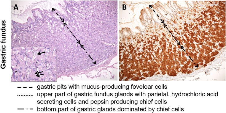
Fig. S4.
Identification of human gastric cell types by H&E staining in gastric fundus showing localization of gastric cell types (A). Parietal cells are localized in the glands of gastric fundus and body and are scattered in the middle and, to a lesser extent, the bottom part of the mucosa. They are characterized by broad pink cytoplasms. Chief cells stain with basophilic cytoplasm and are mainly located in the bottom parts of the mucosa, which can be seen in more detail in the Inset: gastric glands with parietal (single arrow) and chief cells (double arrow). (B) Immunohistochemical localization of taste receptor TASR10.

