Fig. 2.
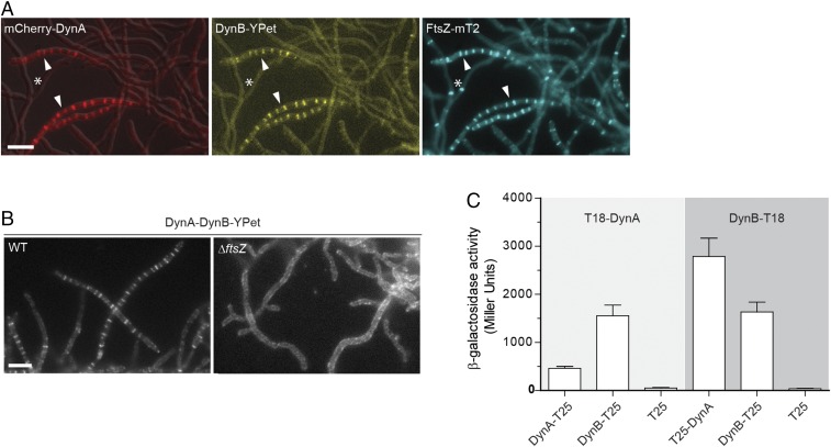
DynA–DynB complexes colocalize with FtsZ at nascent division sites. (A) Subcellular colocalization of fluorescent fusions to DynA (mCherry-DynA) and DynB (DynB-YPet) with FtsZ-mTurquoise2 (FtsZ-mT2). The asterisk denotes vegetative cross-walls and arrowheads point to sporulation septa. Microscopy images of the triply labeled strain (SS206) are representative of at least two independent experiments. (Scale bar: 5 μm.) (B) Localization of DynB-YPet in the WT (SS142) and in the ftsZ null mutant (ΔftsZ, SS238). The dynAB-ypet construct was ectopically expressed from a constitutive promoter (PermE*). (Scale bar: 5 μm.) (C) β-galactosidase activities demonstrating an interaction between DynA and DynB in E. coli BTH101. Positive interaction is detected when DynA and DynB protein fusions to the “T18” and “T25” domains of adenylate cyclase reunite the enzyme, resulting in the synthesis of LacZ. Strains expressing only the T25 domain were used as a negative control. Results are the average of three independent experiments. Error bars represent the SEM.

