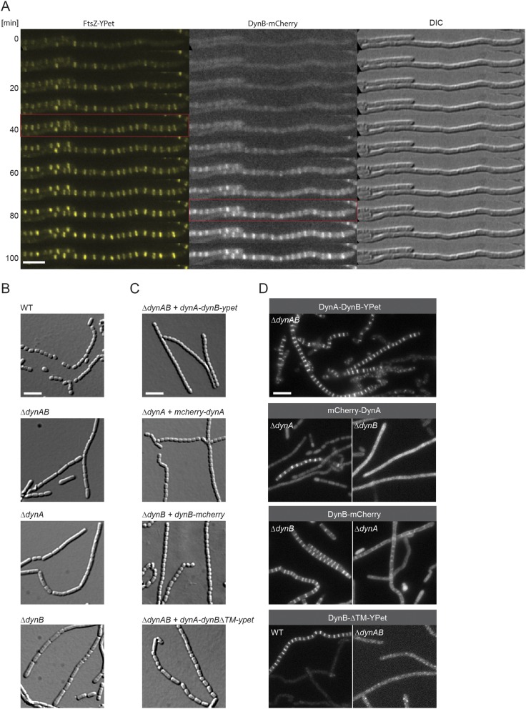
Fig. S2.
DynA and DynB are codependent and function downstream of FtsZ. (A) Montage of a fluorescent time-lapse sequence showing the localization of FtsZ-YPet and DynB-mCherry (SS196). The red box indicates the appearance of FtsZ-ladders (Left) and the colocalization of DynB (Middle). Corresponding DIC images are shown (Right). Imaging interval was 10 min. (Scale bar: 5 μm.) (B) Sporulating aerial hyphae showing the sporulation septation defect in the ΔdynAB (LUV001), ΔdynA (SS255), and ΔdynB (SS2) mutants compared with the WT. Cells were grown for 4 d on MYM and imaged. (Scale bar: 5 μm.) (C) Images of sporulating aerial hyphae showing the ability of the fluorescent dynamin fusions to complement the ΔdynAB phenotype. All constructs were integrated at the ΦBT1 attachment site and expression of the genes was driven either by the dynamin promoter (SS90, SS266) or by the ermE* promoter (SS263, SS89). No complementation was observed when dynBΔTM-ypet was expressed (Bottom, SS263). (Scale bar: 5 μm.) (D) Sporulating aerial hyphae showing the localization codependency of functional fluorescent fusions to full-length DynA or DynB in the ΔdynA (SS266), ΔdynB (SS89, SS190), and ΔdynAB (SS90, SS95) mutants. The fluorescent DynB protein fusion lacking the two transmembrane domains (DynBΔTM-YPet, Bottom) was produced either in the WT (SS262) or the dynamin mutant (SS263). (Scale bar: 5 μm.)

