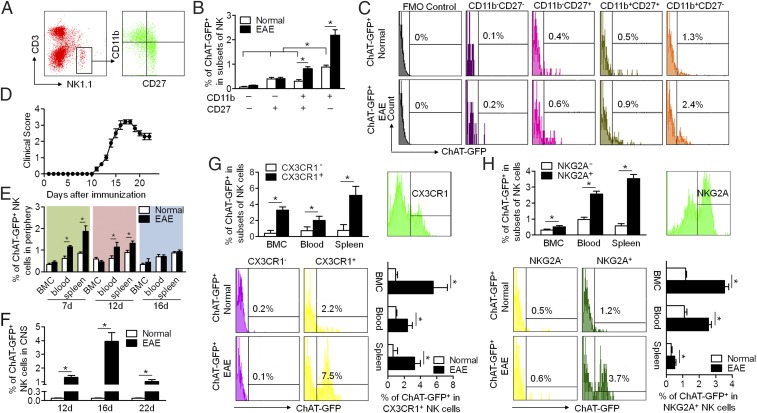
Fig. 2.
ChAT-eGFP expression in murine NK cells during their development and phenotypic maturation upon EAE induction. (A) The development of NK cells is divisible into four stages based on the CD11b expression in combination with CD27. (B and C) ChAT-eGFP expression was determined on subsets of splenic NK cells at different development stages (CD11b−CD27−, CD11b−CD27+, CD11b+CD27+, CD11b+CD27−) in EAE mice. FMO control, fluorescence minus one control. ChAT expression increased during the maturation of NK cells under both normal and EAE conditions. In the presence of EAE, ChAT expression by mature NK cells increased dramatically. n = 6 per group. (D) The disease course of EAE was recorded: Initial manifestations occurred at 11 dpi; the peak was at around 16 dpi; and partial recovery was at 20 to 22 dpi. n = 6 per group. (E) During the course of EAE, the percentage of ChAT+ NK cells in the periphery (blood and spleen) changed dynamically with disease progression, peaked at 7 dpi, recovered slightly at 12 dpi, and returned to normal at 16 dpi. During that time, ChAT-eGFP expression remained constant in naïve mice. n = 6 per group. (F) The percentage of ChAT+ NK cells that infiltrated into the CNS reached a peak at 16 dpi and fell at 20 dpi in EAE mice. n = 12 per group. (G) Circulatory NK cells were divided phenotypically into subsets based on CX3CR1 expression. The expression of ChAT-eGFP in CX3CR1+ NK cells exceeded that of CX3CR1− NK cells. The EAE status increased ChAT-eGFP expression in CX3CR1+ NK cells. n = 6 per group. (H) Functional NK cells from the CNS were divided phenotypically into subsets based on NKG2A expression. The expression of ChAT-eGFP in NKG2A+ NK cells surpassed that of NKG2A− NK cells. The EAE disease state increased ChAT-eGFP expression in NKG2A+ NK cells. n = 6 per group from three independent experiments. Mean ± SEM. *P < 0.05.

