Figure 1.
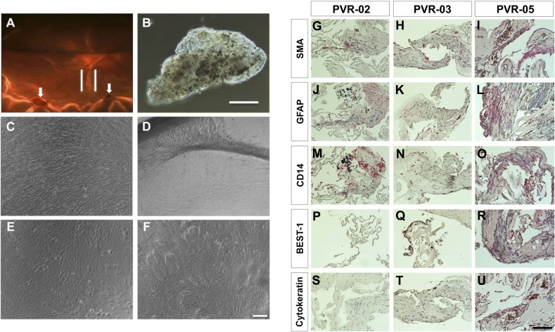
Culture of human PVR membranes and histopathology of PVR membranes. (A) Fundus photograph of the left eye of a patient, case 6 (PVR-06) with recurrent retinal detachment; note presence of a gas bubble within the eye from previous retinal surgery. There is a band of pigmented PVR along the inferior arcade (outlined by white lines). Also note inferior retinal holes in area of detached retina (white arrows). (B) Phase contrast view of the PVR membrane after surgical excision from PVR-06. Scale bar denotes 500 μm. (C) C-PVR cells from case 3 (PVR-03) after 1 week in culture. (D) After 4 weeks in culture, note the formation of bands between the membrane and the rim of the culture dish. (E) C-PVR cells from case 5 (PVR-05) were similarly confluent after 1 week. (F) After 4 weeks in culture, cells grew on top of each other with loss of cell contact inhibition. Scale bar denotes 100 μm. (G–U) Light micrographs of PVR membranes from three different cases (PVR-02, PVR-03, and PVR-05) using primary antibodies (all in red) against SMA (G–I), GFAP (J–L), CD14 (M–O), BEST-1 (P–R), cytokeratin (S–U), and counterstained with hematoxylin (blue). Scale bar denotes 100 μm.

