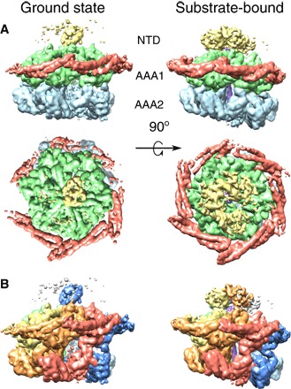
Fig. 1. Domain and protomer organization of ClpB.

(A) Side and top views of the ClpB maps in the ground (left) and substrate-bound (right) states. Maps are segmented and colored according to the domain organization of ClpB protomers: NTD (yellow), MD (red), AAA1 (green), and AAA2 (blue). (B) Side views of the ClpB maps in the ground (left) and substrate-bound (right) states, segmented and colored by protomer. The substrate is shown in purple.
