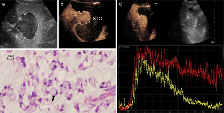
Fig 1. A 65-year-old man with a gastric signet-ring cell carcinoma.
a: Oral contrast agent ultrasound detected an irregular mass in gastric fundus. b: DCUS showed a homogeneous hyper-enhancement tumor. c: Many signet-ring cells (hematoxylin and eosin, × 200) in microscopic field. d: Time-intensity curve depicted the lesion (red line) have faster AT, higher PI and AUC than surrounding normal tissue (yellow).

