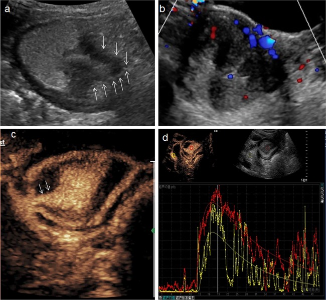
Fig 4. A 65-year-old man with a polypoid adenoma.
a: Conventional trans-abdominal ultrasound with oral contrast agent showed an irregular hypoechoic lesion (arrow) located in antrum. b: Multiple flow signals were visible on the color Doppler. c: DCUS showed homogeneous hyper-enhanced mass connected with gastric wall by a pedicle (arrow). d: Time-intensity curve depicted the lesion (red line) have higher PI than surrounding normal tissue (yellow).

