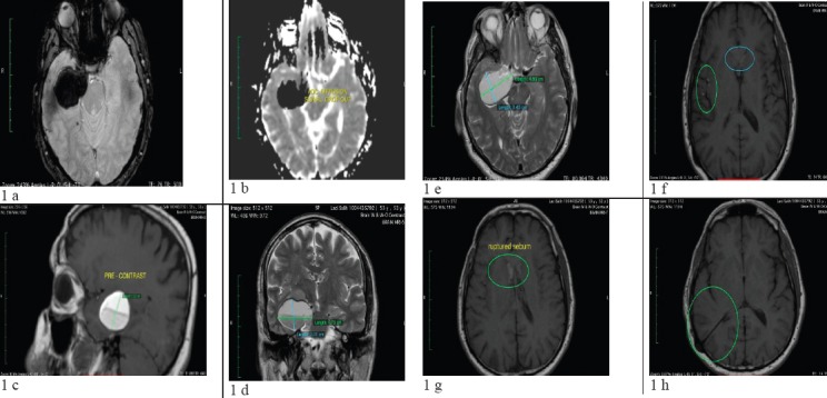
Figure 1.

(a- h): Subsequent MRI of the brain revealed an oval and lobulated 47x34x30mm (TRxAPxCC) non-enhancing T1-hyperintense mass in right cavernous sinus, with compression of surrounding mesial temporal lobe and right anterolateral aspect of mesencephalon. Findings are consistent with ruptured dermoid cyst, given the evacuated sebum content at its lower half. Sebum particles in millimetric sizes are seen within right Sylvian fissure, anterior horns of lateral ventricles and to a lesser extent within left Sylvian fissure, right parietal sulci, cerebral aqueduct, and basal cisterns. No restricted diffusion is seen, eliminating the possibility of epidermoid. A shunt catheter is evident traversing between right lateral ventricle and right parietal bone; besides, slit-like right lateral ventricle is noted (likely secondary to over-draining shunt catheter)
