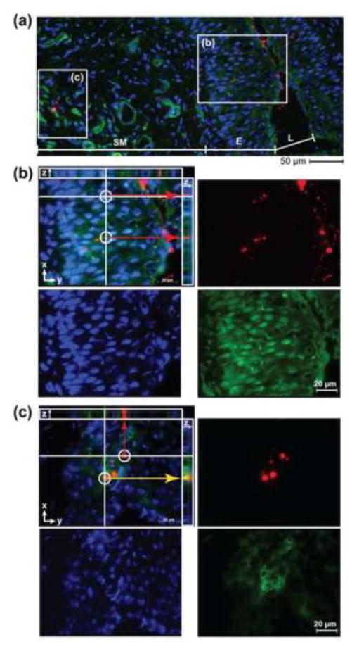
Figure 3. Chitosan promotes translocation of NPs across the vaginal epithelium.
Fluorescent images of NPs (red) merged with IFHC-stained cell nucleus (DAPI, blue) and cytoskeleton (Phalloidin, green) from vaginal tissue sections excised at 24 h following treatment with CH200NP. (A) Image (20× objective) of FRT tissue section demarcating a region of the vaginal epithelium and submucosa. (B) Inset (b) of vaginal epithelium (63× oil objective) from (A) showing an optical section where NPs (red) have penetrated beneath the epithelium. (C) Inset (c) of submucosa (63× oil objective) from (A) showing an optical section where NPs (red) have reached the lamina propria and co-localize (yellow arrows) with cells (green). Images are representative of n = 3 mice.

