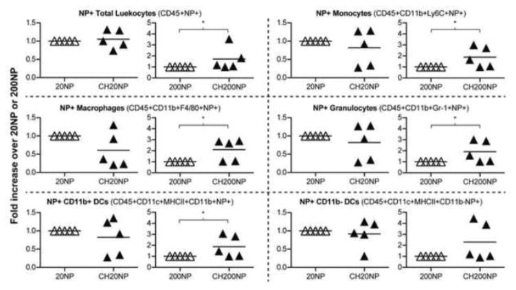
Figure 4. NPs associate with various immune cells in mouse RTs.
Fold-increase in mean fluorescence intensity (MFI) of NP+ cells resulting from chitosan pre-treatment relative to no pre-treatment. Reproductive tracts were excised 24 h post-administration of NPs and isolated cells were stained with fluorescently labeled antibodies to identify specific immune cells under flow cytometry. Data show mean ± SD from n = 5 independent trials (2-4 mice combined per treatment per trial). *: P ≤ 0.05.

