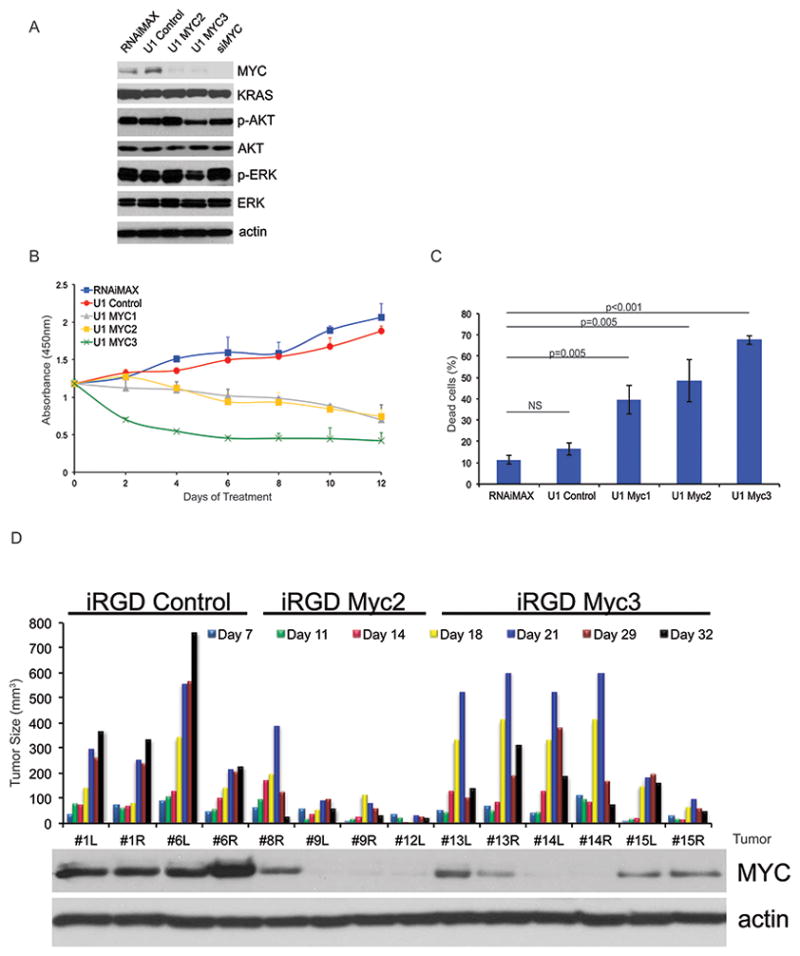
Figure 4. U1 Adaptors targeting MYC in mutant KRAS driven pancreatic cells are effective both in vitro and in vivo.

A, Western blotting of MIA PaCa-2 cells transfected with 10 μM U1 Control, U1 MYC1, U1 MYC2, U1 MYC3, and siMYC after 72 hours of treatment. B, Cell proliferation assay of MIA PaCa-2 cells treated as in A, except for every 72 hours over 9 days. C, Annexin V assay of MIA PaCa-2 cells treated as in A, except cells were harvested on Day 6 following 2 transfections 72 hours apart. Statistical significance was calculated by student’s t-test. D, Nude mice harboring MIA PaCa-2 xenograft tumors were treated 2 times a week for a total of 32 days with 30 μg iRGD-Control (n=4), iRGD-MYC2 (n=4), and iRGD-MYC3 adaptors (n=6). Waterfall plot of tumor size with the correlate protein expression was measured for treated tumors. Western blotting using antibodies for actin and MYC were completed on tumor cell lysates from iRGD-Control, iRGD-MYC2, and iRGD-MYC3 tumors at the end of treatment. Representative images are shown. NS, not significant.
