
Figure 4.
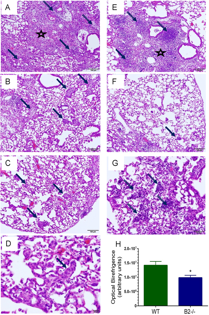
Different histopathological features in WT and B2−/− lungs after clearance of IAV infection. Histopathology was assessed 14 days after infection from two independent experiments. Images shown are representative of n = 7 WT mice and n = 5 B2−/− mice. (A) Fibrotic lesions (star) with areas of metaplasia (solid arrows) and disperse mononuclear infiltrates (lined arrows) consolidate the peribronchial and alveolar spaces of WT mice. (B–D) Cryprogenic organizing pneumonia with buds of granulation tissue in alveoli of WT mice (solid arrows). (E) Fibrotic lesions (star) with areas of metaplasia (solid arrow) and lymphocytic foci (lined arrows) consolidate the peribronchial and alveolar spaces of B2−/− mice. (F) Mild (solid arrow) to absent cryptogenic organizing pneumonia in B2 alveoli. (G) Alveolar lymphocytic infiltrates in B2−/− lungs. Magnification for A, B, C, E, and F: 100x; D: 600 x; G: 400x. (H) Comparison of collagen birefringence in WT and B2−/− lungs. Collagen was visualized picric sirius red stain. *p < 0.00046. Data shown are means ± SEM from total of 50 and 60 microscopic fields at 200x magnification in lung sections from n = 4 WT mice and n = 5 B2−/− mice, respectively.