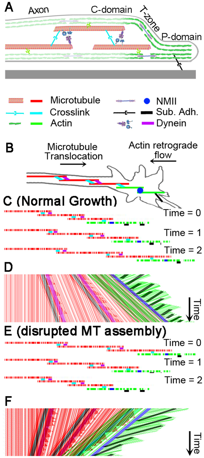
Figure 5.

Model of neurite elongation by bulk forward translocation of MTs. (A) Side view of growth cone and distal neurite with important cytoskeletal elements. (B) Schematic of a growth cone and distal neurite with important cytoskeletal components and substrate adhesion (black). (C) Three MTs (red) and two actin filaments (green) labeled with fluorescent speckles at different time points. (D) Hypothetical kymograph of MTs (red), F-actin (green), motors (purple and blue), and cross linking proteins (light blue). Anterograde red lines reveal forward movement of MTs in the distal neurite and C-domain and retrograde green lines reveal retrograde movement of F-actin in P domain. (E) Illustration of MT and F-actin configuration under conditions where disruption of MT assembly induces retraction of the growth cone. (F) A hypothetical kymograph of MT and F-actin motion. While axonal frictional interactions with the substrate, as well as axonal actin and myosin activity are all important for both elongation and retraction, they are not shown to minimize the complexity of the figure.
