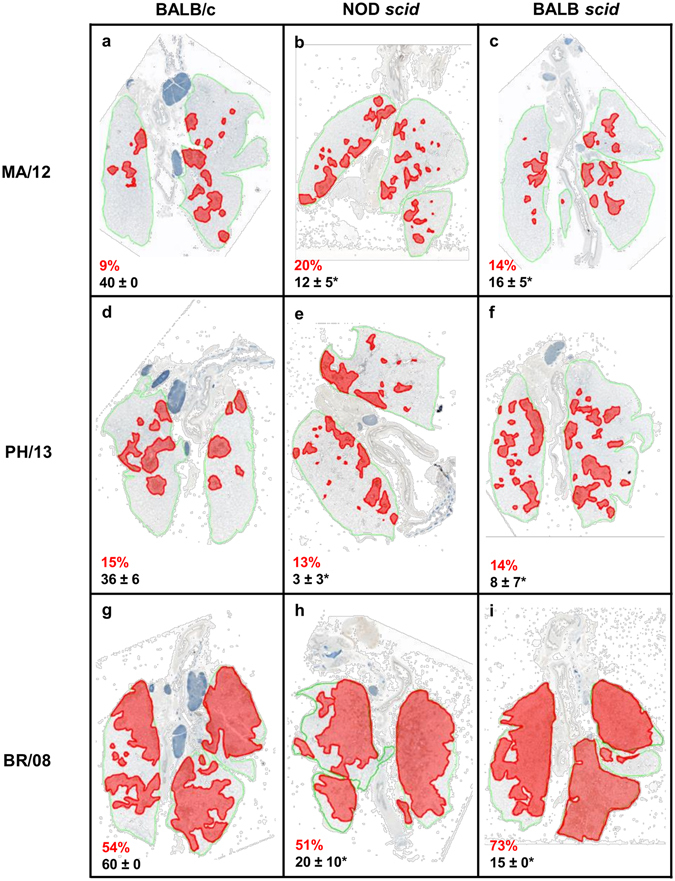
Figure 4.

Histopathologic changes in the lungs of immunocompetent and immunocompromised mice inoculated with influenza B viruses. BALB/c, NOD scid, and BALB scid mice were inoculated with viruses as described in the legend for Fig. 2. Pulmonary lesions were evaluated at 6 dpi (n = 4/virus/mouse strain). The panels show the extent of virus infection and histopathology of influenza MA/12, PH/13, and BR/08 viruses in the lungs of immunocompetent BALB/c (a,d,g) and immunocompromised NOD scid (b,e,h) and BALB scid (c,f,i) mice. A representative section is shown for each virus and mouse strain. The green lines designate total lung areas measured; red-shaded areas designate bronchioles/alveoli with active virus infection (i.e., containing antigen-positive epithelial cells). The average percentage of the total lung field containing antigen-positive epithelial cells are shown in red texts and the average severity score ± SE for inflammation are shown in black texts. *P < 0.05, relative to BR/08 scores, as determined by one-way ANOVA with Bonferroni’s multiple comparison post-test.
