Figure 2.

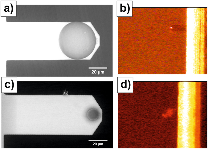
Role of probe size in the interaction with mucus blankets. (a) Representative SEM image of the 40 µm colloidal probes used in this study (the actual diameters were always determined from the analysis of the SEM images). (b) Optical image registered when the probe shown in (a) established contact with ileum mucosa at a repulsive force of 10 nN. (c) Representative SEM image of the 15 µm colloidal probes used in the study. (d) Optical image registered when, while approaching ileum mucosa with the probe shown in (c), the signal registered by the PSD dropped to zero.
