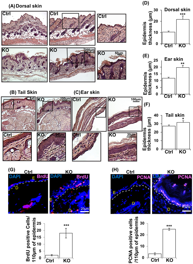
Figure 2.

Loss of N-WASP expression in keratinocytes caused hyperproliferation of keratinocytes in N-WASPK14KO mice. (A) H & E stained dorsal skin, tail (B) and ear (C) sections of N-WASPK14KO and control mice on 23rd day (n = 10). Quantification of epidermal thickening in skin (D), ear (E) and tail (F) of control mice and N-WASPK14KO mouse skin sections (n = 5). (G) BrdU staining: Quantification of BrdU positive cells in epidermis of control (n = 3) and N-WASPK14KO (n = 4) mice skin at P23 (H) PCNA immunostaining of paraffin embedded dorsal skin sections of control and N-WASPK14KO mice at P23 and quantification of PCNA positive cells (n = 3). Results are mean ± SEM ***p < 0.001, **p < 0.01, *p < 0.05.
