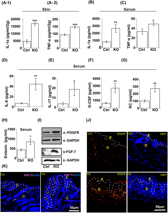
Figure 8.

Development of local and systemic inflammatory responses in N-WASPK14KO mice. Cytokine analysis showed higher levels of IL-1α (A-1) and TNF-a (A-2) in skin and as well as in serum (B and C), IL-6 (D) and IL-17 (E) cytokines in serum of N-WASPK14KO compared to control mice on 23rd day. Similarly, chemokine analysis showed significantly increased levels of G-CSF (F), KC (G) and eotaxin (H) chemokines in serum of N-WASPK14KO mice compared to control mice on 23rd day. (I) Western blot analysis of whole skin protein lysate showed that expression of PDGFR and FGF-7 are increased in N-WASPK14KO compared to P23 control mice (Blots were cropped and full length western blot images are in supplementary Fig. S6C). Immunostaining showed that both PDGFR (J) and FGF-7 (K) are significantly increased in skin of N-WASPK14KO mice compared to control mice (P23 mice). Results are mean ± SEM ***p < 0.001, **p < 0.01, *p < 0.05 (n = 8).
