
Figure 4.
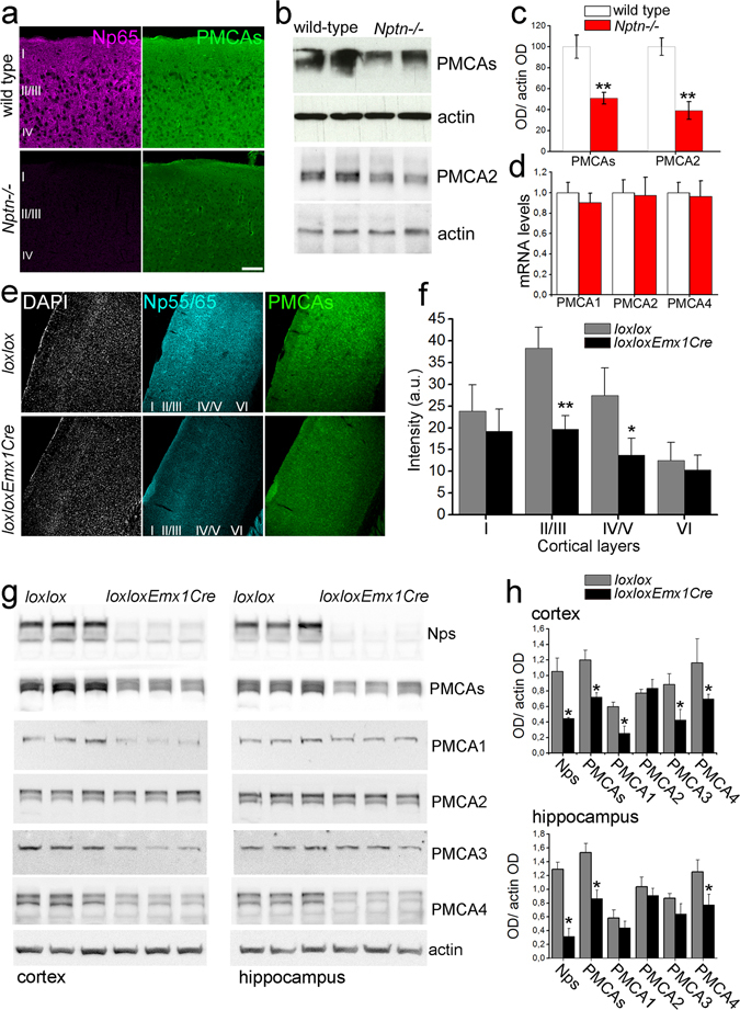
Distinct loss of PMCA isoforms in Nptn loxloxEmx1Cre brain. Reduction in total PMCA immunoreactivity was assessed using (a) Np65 and pan-PMCAs antibodies and confocal microscopy in Nptn −/− cortical slices (scale bar = 100 µm) or (b) pan-PMCAs and PMCA2 antibodies and Western blot analysis of homogenates from Nptn −/− brains. (c) Intensity quantification of the Western blots in b. Six wild-type (white bars) and six Nptn −/− (red bars) independent samples were quantified. (d) Quantitative RT-PCR for PMCA1, 2, and 4 in Nptn −/− brain samples. PMCA products were normalized to control GAPDH products and then, each individual mutant was normalized to the corresponding wild-type paralog value. (e) Representative confocal sections from Nptn loxloxEmx1Cre and Nptn loxlox somatosensory cortex stained with a pan-Np55/65 (cyan) and a pan-PMCA antibody (green) with DAPI as counterstain (grey). (f) Quantification of PMCA staining intensity throughout cortical layers of Nptn loxlox (gray bars) and Nptn loxloxEmx1Cre (black bars) mice (three confocal sections per animal, 4 animals per genotype were quantified as described)10, 13. (g) Homogenates from cortex and hippocampus of Nptn loxloxEmx1Cre (loxloxEmx1Cre) and Nptn loxlox (loxlox) mice were analyzed by Western blot using paralog-specific PMCA and pan-Np55/65 antibodies as indicated. Actin was used as loading control. (h) Intensity quantification of the Western blots in g. Three Nptn loxlox (gray bars) and three Nptn loxloxEmx1Cre (black bars) independent samples were quantified. Data are mean ± SEM with *P < 0.05 or **P < 0.01, unpaired two-tailed t-test. For confocal images, cortical layers are indicated.