
Figure 3.
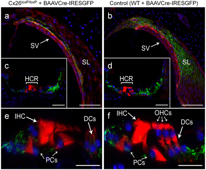
Confocal immunofluorescence imaging of cochlear cross–sections from mice injected at P25 with BAAVCre–IRESGFP viral vectors. Color code: Cx26, green; actin filaments, red; nuclei, blue. (a,b) Stria vascularis (SV) and spiral ligament (SL). (c,d) organ of Corti; HCR, hair cell region. (e,f) close–up view of the HCR from c,d, respectively. DCs: Deiters’ cells; IHC: inner hair cell; OHCs: outer hair cells; PCs: pillar cells. Scale bars: 50 μm in (a–d); 25 μm in (e,f).