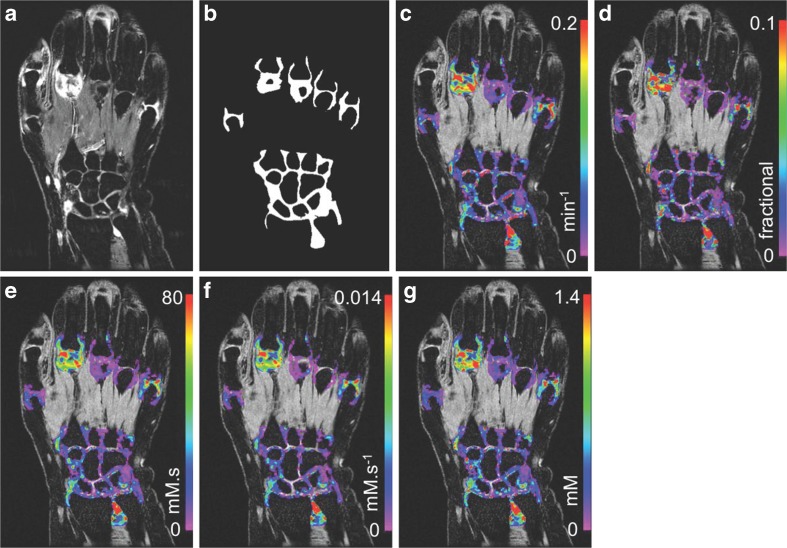
Fig. 1.
Post-contrast high resolution T 1-weighted spoiled gradient-recalled echo (SPGR) image with fat saturation shows predominant disease in the second metacarpal joint (MCP) and isolated areas of disease in MCP-5, the distal radio-ulnar and radio-carpal joints (a). Segmented joint voxel masks were produced for each joint and used in the DCE-MRI analysis (b). Pre-contrast images with DCE-MRI parameterisation overlays for: K trans (c), v p (d), IAUC 120 (e), IRE (f) and ME (g)

