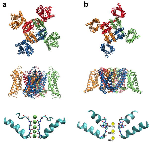
Figure 1.
Overall structure of voltage-gated K+ (Kv) and Na+ (Nav) channels. The Kv channel (a) is the mammalian Kv1.2/Kv2.1 chimera structure protein data base (PDB) entry: 2R9R (Long et al., 2007). The Nav channel (b) is the bacterial NavAb structure PDB entry 3RVY (Payandeh et al., 2011). At the top is a view of the tetrameric channels seen from the extracellular side. In the middle is a side view of the channels. At the bottom is a close-up view of the selectivity filter. In the Kv channel (a), the narrow selectivity filter of K+ channels comprises 5 distinct binding sites (S0 to S4) where the permeating K+ ions are coordinated by backbone carbonyl oxygens. In the Nav channel (b), the selectivity filter is relatively wide, allowing the passage of partially hydrated ions. A few binding sites have also been identified (Payandeh et al., 2011).

